parts of microscope and functions pdf
Microscopes are essential tools, magnifying small objects for detailed examination. Understanding their parts is crucial for effective use. This includes structural, optical, and mechanical components. Each part plays a vital role in achieving magnification and clear imaging.
The Importance of Microscopes
Microscopes are indispensable in numerous fields, from biology to materials science, enabling the visualization of structures invisible to the naked eye. They allow scientists to explore cellular components, microorganisms, and tissue samples, advancing medical research and diagnostics. In material science, microscopes help analyze material properties at a microscopic level. Their ability to magnify and reveal intricate details is fundamental for scientific discovery, quality control, and technological innovation. Furthermore, microscopes play a vital role in education, allowing students to observe and learn about the microscopic world. By providing a window into the unseen, these instruments facilitate a deeper understanding of the world around us.

Structural Components of a Microscope
The structural components of a microscope provide the framework for its operation. These include the head, arm, and base. They ensure stability and support for the optical and mechanical elements.
Head (Body Tube)
The head, also known as the body tube, is a crucial component of the microscope’s structure. It is essentially a cylindrical, often metallic, tube. This tube serves as the main housing for the optical elements in the upper portion of the microscope. At one end, the head connects to the eyepiece lens, which is the lens closest to the viewer’s eye, and at the other end, it connects to the nosepiece, which holds the objective lenses. The body tube ensures correct alignment and distance between the eyepiece and the objective lenses. This precise alignment is essential for creating a clear and focused image of the specimen. The head’s primary function is to maintain the proper optical path within the microscope, and it helps in achieving the desired magnification.
Arm
The arm of a microscope is a vital structural component that connects the head of the microscope to its base. Functionally, it acts as a supportive frame, linking the optical components to the foundation of the microscope. This connection is crucial for stability and precise positioning of the optical elements. The arm provides a secure grip when carrying the microscope, ensuring the delicate parts are not damaged during transport. It also helps maintain the proper distance between the optical and mechanical components of the instrument. The arm is designed to be robust, providing a solid and stable framework for the entire microscope, which is essential for accurate observations. Proper support from the arm allows for easy adjustments during use.
Base
The base of a microscope is the foundational structure providing stability and support for the entire instrument. It is typically a wide, flat platform that rests on the working surface, ensuring the microscope remains steady during use. The base plays a critical role in preventing the microscope from tipping over, especially when adjustments are being made. Often, the base contains the illuminator, which provides the light source for viewing specimens, and the controls for adjusting the light intensity. Additionally, it provides a convenient location for the on/off switch and other electrical connections. The base’s design contributes to the overall ergonomic structure of the microscope, promoting ease of use and comfortable operation. Its weight is an important factor in its stability.
Optical Parts of a Microscope
The optical parts are essential for magnification and image formation. These include lenses like the eyepiece, objective lenses, and the condenser, which work together to provide a clear and enlarged view of the specimen.
Eyepiece (Ocular Lens)
The eyepiece, also known as the ocular lens, is the lens located at the top of the microscope that the user looks through. It typically provides a magnification of 10x or 15x, further enlarging the image created by the objective lens. This lens is crucial for viewing the magnified specimen, and it is part of the optical system. The eyepiece works in conjunction with the objective lens to produce the final image, and it is positioned at the upper end of the body tube. It is an essential component for comfortable viewing and detailed observation. The eyepiece lens is designed to provide a clear and focused image, ensuring the user can see the magnified specimen clearly. It is responsible for the final magnification step before the image reaches the observer’s eye, allowing for clear and detailed examination of specimens under the microscope.
Objective Lenses
Objective lenses are crucial components of a microscope, situated closest to the specimen being observed. They are responsible for the initial magnification of the specimen. These lenses are mounted on a rotating nosepiece, allowing the user to select different magnifications, typically ranging from 4x to 100x. Each objective lens has a specific magnification power and numerical aperture, influencing the resolution and clarity of the image. The objective lens collects light passing through the specimen and creates a magnified, real image that is then further magnified by the eyepiece. Different objective lenses are used for observing specimens at different scales and with varying levels of detail. Choosing the correct objective lens is essential to obtaining high-quality images of the specimen for accurate analysis. They are key to resolving fine details within specimens.
Condenser
The condenser is a crucial optical component located beneath the microscope stage. Its primary function is to focus and concentrate the light from the illuminator onto the specimen. This focused light is essential for achieving optimal illumination and contrast for clear viewing. The condenser typically consists of a series of lenses that gather light and direct it through the specimen. It can be adjusted vertically to control the intensity and focus of light, allowing users to tailor the illumination to the specific specimen and objective lens being used. A well-adjusted condenser is critical for obtaining high-quality images with good resolution and contrast. The condenser allows the user to optimize the lighting for various magnifications and specimen types. It is an essential component for proper image formation.

Mechanical Parts of a Microscope
Mechanical parts provide support and enable adjustments for clear viewing. They include the stage, nosepiece, and focusing knobs, ensuring precise movement and stability during specimen observation.
Stage
The stage is a flat platform where the microscope slide containing the specimen is placed for observation. It provides a stable surface to hold the slide securely during viewing and prevents it from moving unintentionally. Some stages come equipped with stage clips to further secure the slide, especially when using higher magnifications. Many stages also have mechanical adjustments that allow for precise movement of the slide on the x and y axes, enabling the user to view different parts of the specimen. These adjustments allow for a systematic scanning of the slide and are especially useful for observing larger specimens or multiple fields of view. The stage is a key component in any microscope and is essential for proper specimen placement and examination.
Nosepiece
The nosepiece, also referred to as the revolving nosepiece, is a critical component of a compound microscope that holds multiple objective lenses. These lenses vary in their magnification power, allowing the user to switch between different levels of magnification. The nosepiece rotates, enabling the user to easily select the appropriate objective lens for the desired level of detail when observing a specimen. This is a vital feature for any multi-lens microscope, providing flexibility in viewing at different scales. The nosepiece ensures that the selected objective lens is aligned with the optical path for proper image formation. It contributes significantly to the overall functionality and versatility of the microscope, making it a valuable component for users.
Focusing Knobs (Coarse and Fine)
Focusing knobs are essential for achieving a clear image under a microscope. They come in two types⁚ coarse and fine focus knobs. The coarse focus knob provides large adjustments, rapidly moving the stage or the objective lens to bring the specimen into approximate focus. This knob is used first when initially focusing on a slide. The fine focus knob, on the other hand, offers minute adjustments, allowing for precise focusing to sharpen the image. This knob is used after the coarse focus, ensuring the image is as clear and detailed as possible. Both knobs work in conjunction to enable accurate and sharp observation of specimens. Therefore, they are crucial for obtaining clear microscopic images.

Other Important Microscope Parts
Beyond the main components, the illuminator provides light for viewing, and the diaphragm adjusts the light intensity. These parts are critical for achieving optimal image clarity and contrast.
Illuminator
The illuminator, a crucial component of any microscope, is the light source that provides the necessary illumination for observing specimens. This component can be a simple mirror that reflects ambient light, or, more commonly, an integrated electric lamp. The illuminator’s primary function is to direct light through the specimen on the stage, ensuring that it is adequately illuminated for viewing. The intensity of light from the illuminator is often adjustable, allowing users to tailor the brightness to suit the specific requirements of the specimen and magnification being used. Proper illumination is essential for achieving clear, high-contrast images and for accurate observation of minute details. Without an effective illuminator, the specimen would appear dark, indistinct, and difficult to analyze. The position and type of the illuminator are also important factors that affect the quality of the image.
Diaphragm
The diaphragm, an essential part of the microscope, is located beneath the stage and plays a key role in controlling the amount of light that passes through the specimen. It functions like an iris in a camera, adjusting the diameter of the light beam that reaches the condenser and subsequently the slide. This control is crucial for optimizing image clarity and contrast. A fully open diaphragm allows maximum light to pass, which can be useful for low-magnification viewing. Conversely, closing the diaphragm reduces the amount of light, enhancing contrast at higher magnifications. Proper adjustment of the diaphragm helps prevent over-illumination, which can wash out detail, and under-illumination, which makes it difficult to see the specimen. Mastering the diaphragm’s usage is vital for any microscopist, ensuring optimal viewing conditions and enabling clear observation of the specimen’s features.

Types of Microscopes
Microscopes vary widely, including simple, using single lenses, and compound, employing multiple lenses for higher magnification. Other types exist, each designed for specific applications and offering unique advantages in scientific study.
Simple Microscope
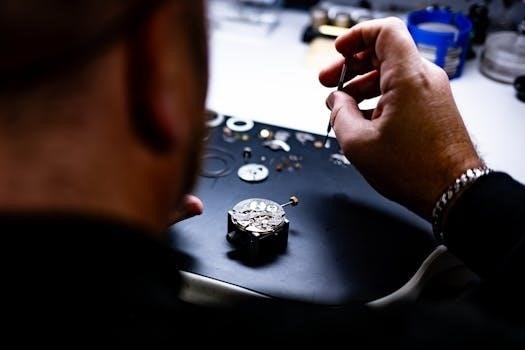
A simple microscope, characterized by its use of a single convex lens, provides basic magnification for viewing small objects. This type of microscope is foundational in the history of microscopy, offering a straightforward way to enlarge images. Often employed in contexts such as watchmaking, jewelry examination, and detailed work by miniature artists, it allows for close inspection of fine details. Its uncomplicated structure makes it a useful tool for basic observations. While limited in magnification compared to compound microscopes, the simple microscope continues to hold relevance for specific applications, demonstrating that effective magnification can be achieved with minimal complexity, it is used for industrial applications and more.
Compound Microscope
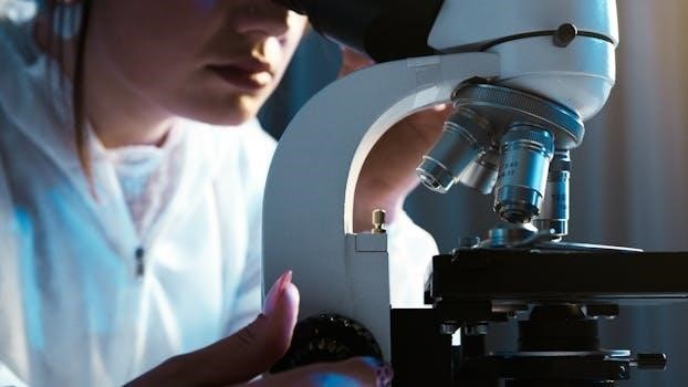
The compound microscope, unlike its simple counterpart, employs multiple lenses to achieve higher magnification. It utilizes an objective lens close to the specimen and an eyepiece lens near the viewer’s eye, this system significantly enhances the magnification and resolution of minute details. This advanced design makes it an indispensable tool for scientific research, medical diagnostics, and educational purposes. The compound microscope allows for detailed observation of cells, bacteria, and other microorganisms, revealing complex structures that would be invisible to the naked eye. Its intricate system of lenses and mechanical adjustments provides a powerful means for exploring the microscopic world, with varying configurations.
